CHAPTER 70 CLASSIFICATION AND CLINICAL MANIFESTATIONS OF NEUTROPHIL DISORDERS
MARSHALL A. LICHTMAN
Classification
Clinical Manifestations
Neutropenia
Qualitative Neutrophil Abnormalities
Neutrophilia
Neutrophil-Induced Vascular or Tissue Damage
Chapter References
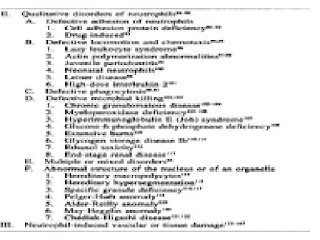
Neutrophil disorders can be grouped into deficiencies, or neutropenia, and excesses, or neutrophilia. The former can have the severe consequence of predisposing to infection, whereas the latter is usually a manifestation of an underlying inflammatory or neoplastic disease: the neutrophilia, per se, having no specific consequences. Neutropenia may reflect an inherited disease that is usually evident in childhood (such as congenital neutropenia), but it is more often acquired. The most common cause for neutropenia is the adverse effect of the use of a drug. Some cases of neutropenia have no evident cause. The health consequence of neutropenia is a function of the severity of the decrease in the blood neutrophil count and the abruptness and duration of the decrease. Qualitative disorders of neutrophils may lead to infection as a result of defective chemotaxis to an inflammatory site or defective microbial killing. Table 70-1 provides a comprehensive categorization of neutrophil disorders.
TABLE 70-1 CLASSIFICATION OF NEUTROPHIL DISORDERS
CLASSIFICATION
Table 70-1 lists disorders that result from a primary deficiency in neutrophil numbers or function. Neutropenia or neutrophilia may also occur as part of a disorder that affects multiple blood cell lineages, such as occurs in infiltrative diseases of the marrow or after cytotoxic drug therapy, but these are not included in this classification and are discussed in other parts of this text. In this classification, and in this section of the text, we consider diseases resulting from neutrophil deficiencies in which the neutrophil is either the only cell type affected or is the dominant cell type affected.
A pathophysiologic classification of neutrophil disorders has proved elusive. Techniques to measure mechanisms of impaired production or accelerated destruction of neutrophils are more difficult and complex than those used for red cells or platelets. The low concentration of blood neutrophils, accentuated in neutropenic states, makes radioactive labeling techniques to study the kinetics of autologous cells in neutropenic subjects technically difficult or not possible. The two compartments of neutrophils in the blood, the random disappearance of neutrophils from the circulation, the extremely short circulation time of neutrophils, the absence of techniques to measure the size of the tissue neutrophil compartment, and the disappearance of neutrophils by death or excretion from the tissue compartment also make multicompartment kinetic analysis exceedingly difficult. Also, neutropenic disorders are uncommon, and few laboratories are able, or prepared, to undertake the studies necessary to define the mechanisms of their development in sporadic cases. Therefore, efforts to understand the pathophysiology of neutropenia have had limited success. Hence, the classification of neutrophil disorders is partly pathophysiologic and partly descriptive (see Table 70-1). Although imperfect, classification does provide a language for communication and a basis for rectification as knowledge of the cause and mechanism of disease advances.
The classification is self-explanatory except in two areas. First, certain childhood syndromes have been listed under decreased neutrophilic granulopoiesis. They could have been listed under chronic hypoplastic or chronic idiopathic neutropenia; however, they seem to hold a special interest, and their pathogenesis is still disputed. Three childhood syndromes, although associated with neutropenia, are omitted because the neutropenia is part of a more global suppression of hemopoiesis: Pearson syndrome,1,2 Fanconi syndrome,3,4 and dyskeratosis congenita.5,6
A second area requiring explanation is the chronic idiopathic neutropenias. This group includes: (1) cases with normocellular marrows but an inadequate compensatory increase in granulopoiesis for the degree of neutropenia and (2) cases with hyperplastic granulopoiesis that is apparently ineffective. Unlike hypoplastic neutropenias in which the granulocyte precursors are markedly reduced or absent, precursors are present in the marrow in the idiopathic neutropenias, but the extent of effective granulopoiesis is probably low.
Qualitative disorders of neutrophils affect their ability to enter inflammatory exudates, to ingest microorganisms, or to kill ingested microorganisms (see Chap. 72).
CLINICAL MANIFESTATIONS
The clinical manifestations of decreased concentrations or abnormal function of neutrophils are principally the result of infection.
The combined deficit of neutrophils and monocytes characteristic of aplastic anemia, hairy-cell leukemia, and cytotoxic therapy leads to susceptibility to a broader spectrum of infectious agents. Increased concentrations of normal neutrophils per se have not been associated with clinical manifestations, although increased concentrations of leukemic neutrophil precursors can produce clinical manifestations of microcirculatory leukostasis (see Chap. 91).
NEUTROPENIA
The lower limit of the normal neutrophil count is about 1800/µl (1.8 × 10⁹/liter) in subjects of European descent and 1400/µl (1.4 × 10⁹/liter) in subjects of African descent.148,149,150,151,152,153 and 154 This finding is especially striking in Yemenite Jews, another ethnic group with very low “normal” neutrophil counts.155 A decrement in neutrophil concentration to 1000/µl (1.0 × 10⁹/liter) usually poses little threat in the otherwise healthy individual. If the neutrophil count drops further, the risk of infection increases, and subjects chronically neutropenic as a result of a production abnormality with counts less than 500 neutrophils/µl (0.5 × 10⁹/liter) are at risk of developing recurrent infections.156
The relationship of frequency or type of infection to neutrophil concentration is an imperfect one. The cause of the neutropenia, the coincidence of monocytopenia or lymphopenia, concurrent use of alcohol or glucocorticoids, and other factors can influence the likelihood of infection.
Infections in neutropenic subjects, not otherwise compromised, are most likely to result from gram-positive cocci and usually are superficial, involving skin, oropharynx, bronchi, anal canal, or vagina. However, any site may become infected, and gram-negative organisms, viruses, or opportunistic organisms may be involved.
A decrease in neutrophil count can occur abruptly or gradually (see Chap. 71). One type of drug-induced neutropenia is distinguished by the rapidity of onset. This abrupt-onset neutropenia is more likely to be severe and lead to symptoms. If the neutrophil count approaches zero (agranulocytosis), high fever; chills; necrotizing, painful oral ulcers (agranulocytic angina); and prostration may occur, presumably as a result of sepsis.157,158 and 159 As the disease progresses, headache, stupor, and rash may develop. In the preantibiotic era, persistent agranulocytosis had a fatality rate approaching 100 percent. Even with bactericidal, broad-spectrum antibiotics, severe, sustained neutropenia or agranulocytosis is a serious illness with a high fatality rate.
There is a decrease in the formation of pus in patients with severe neutropenia.160,161 This failure to suppurate can mislead the clinician and delay identification of the site of infection because minimal physical or radiographic findings develop. For example, lack of pneumonic consolidation is characteristic of pneumonia in granulocytopenic subjects. Exudate, swelling, heat, and regional adenopathy are much less prevalent in granulocytopenic patients. Fever is common, and local pain, tenderness, and erythema are nearly always present despite a marked reduction in neutrophils.162,163 and 164
The mechanism of neutropenia, as well as the severity of the deficiency of cells, plays a role in clinical manifestations. Chronic idiopathic (benign) neutropenia is associated with normal granulopoiesis in the marrow and is asymptomatic even when present for prolonged periods, sometimes in the face of neutrophil counts approaching zero.49 Presumably the delivery of neutrophils from marrow to tissues is sufficient to prevent infection despite the low blood pool size.50,51 Monocyte counts are normal, and this may also aid in host defenses, since these cells are effective phagocytes.
Chronic idiopathic (symptomatic) neutropenia is often associated with pyoderma and otitis media in children.55 The former is usually caused by Staphylococcus aureus, Escherichia coli, and Pseudomonas spp., and the latter is usually the result of infection by pneumococci or Pseudomonas aeruginosa. Unexplained chronic gingivitis also may be a manifestation of chronic neutropenia.165 Pneumonia, lung abscesses, stomatitis, hepatic abscesses, or infections in other sites may occur.56
Chronic cyclic neutropenia is characterized by periodic oscillations in the number of neutrophils, with the nadir occurring at about 3-week intervals.36,166 During neutropenia, patients develop malaise, fever, and buccal, labial, or lingual ulcers, and cervical adenopathy. Furuncles, carbuncles, cellulitis, infected cuts with lymphangitis, chronic gingivitis, and abscesses of the axilla or groin also may occur. Although severe infections may lead to fatality, life-threatening complications are uncommon (see Chap. 71).
Some individuals may have neutropenia because a larger proportion of their blood neutrophils is in the marginal rather than in the circulating pool. The total blood neutrophil pool is normal, and infections do not result from this atypical distribution of neutrophils.167 This type of alteration has been called pseudoneutropenia.
QUALITATIVE NEUTROPHIL ABNORMALITIES
Neutrophil function depends on the ability of neutrophils to adhere to endothelium, move, respond to chemotactic gradients, ingest microorganisms, and kill ingested pathogens. Loss of any of these functions can predispose to infection (see Chap. 72). Defects in each step of the neutrophil's participation in the inflammatory response have been identified.168,169 Defects in cytoplasmic contractile proteins, granule synthesis or contents, or intracellular enzymes may underlie a movement, ingestion, or killing defect. These defects may be congenital or acquired. Chronic granulomatous disease102,103 and Chédiak-Higashi disease121 are two examples of the former. Among the acquired disorders are those extrinsic to the cell, such as in the movement, chemotactic, or phagocytic defects of diabetes mellitus,168,169,170 and 171 alcohol abuse,172,173 or glucocorticoid excess.174 Acquired intrinsic disorders are usually manifestations of stem cell disorders like preleukemia175 (see Chap. 91).
Severe defects in bacterial killing, such as occur in chronic granulomatous disease, result in S. aureus, Klebsiella-Aerobacter, E. coli, and other catalase-positive bacterial infections. Suppurative lymphadenitis, pneumonia, dermatitis, hepatic abscesses, osteomyelitis, and stomatitis occur, and chronic granulomatous reactions in these sites give the disease its name. Fatality rates have been high. Functional disorders may be severe, as in chronic granulomatous disease. Mild functional disorders predispose to infections that are relatively infrequent and that respond readily to antibiotics. Severe functional disorders result in suppurative lesions because neutrophil influx into inflammatory foci is not impaired, whereas agranulocytosis is associated with nonsuppurative lesions.
NEUTROPHILIA
An overabundance of neutrophils has not been shown to result in specific clinical manifestations. Neutrophils can transiently occlude capillaries, as determined by supravital microscopy, and such occlusions may reduce local blood flow transiently and contribute to the development of ischemia.124 Impairment of reperfusion of the coronary microcirculation has been thought to be dependent, in part, on neutrophil plugging of myocardial capillaries.123
NEUTROPHIL-INDUCED VASCULAR OR TISSUE DAMAGE
Neutrophil products may contribute to the pathogenesis of inflammatory skin, bowel, synovial, glomerular, and bronchial and interstitial pulmonary diseases.124,125,126,127,128,129,130,131,132,133,134,135,136,137 and 138 In addition, these products may act as mediators of tissue injury in myocardial infarction.139,140,141 and 142 Also, highly reactive oxygen products of neutrophils may be mutagens that increase the risk of neoplasia.144,145 This action may explain, for example, the development of carcinoma of the bowel in patients with chronic ulcerative colitis and the relationship between elevated leukocyte count and the occurrence of lung cancer, independent of the effect of cigarette usage.146 The oxidants, especially hypochlorous acid and chloramines, released by the neutrophil are extremely short lived and may play a role in tissue injury by inactivating several protease inhibitors in tissue fluids, permitting proteases, especially elastase, collagenase, and gelatinase, to cause tissue injury.129 Thrombogenesis has also been ascribed to leukocyte products.143
CHAPTER REFERENCES
1.
Pearson HA, Lobel JS, Kocoshis SA, et al: A new syndrome of refractory sideroblastic anemia with vacuolization of marrow precursors and exocrine pancreatic dysfunction. J Pediatr 95:976, 1979.
2.
van de Corput MP, van den Ouweland JM, Dirks RW, et al: Detection of mitochondrial DNA deletions in human skin fibroblasts of patients with Pearson's syndrome by two-color fluorescence in situ hybridization. J Histochem Cytochem 45:55, 1997.
3.
Gordon-Smith EC, Rutherford TR: Fanconi anemia. Ballières Clin Haematol 2:139, 1989.
4.
d'Appolito M, Zelante L, Savoia A: Molecular basis of Fanconi anemia. Haematologica 83:533, 1998.
5.
Srinavin C, Trowbridge A: Dyskeratosis congenita: Clinical features and genetic aspects. J Med Genet 12:339, 1975.
6.
Dokal I: Severe aplastic anemia including Fanconi's anemia and dyskeratosis congenita. Curr Opin Hematol 3:453, 1996.
7.
Kostmann R: Infantile genetic agranulocytosis. Acta Pediatr Scand 64:362, 1975.
8.
Tidow N, Pilz C, Teichmann B, et al: Clinical relevance of point mutations in the cytoplasmic domain of granulocyte colony-stimulating factor receptor gene in patients with severe congenital neutropenia. Blood 89:2369, 1997.
9.
Rappeport JM, Parkman R, Newburger P, et al: Correction of infantile agranulocytosis (Kostmann's syndrome) by allogeneic transplantation. Am J Med 68:605, 1980.
10.
Kyds U, Pietsch T, Welte K: Expression of receptors for granulocyte colony-stimulating factor on neutrophils from patients with severe congenital neutropenia and cyclic neutropenia. Blood 79:1144, 1992.
11.
Bonilla MA, Gillio AP, Ruggeiro M, et al: Effects of recombinant human granulocyte colony-stimulating factor on neutropenia in patients with congenital agranulocytosis. N Engl J Med 320:1574, 1989.
12.
Weston B, Todd RF III, Axtell R, et al: Severe congenital neutropenia: Clinical effects and neutrophil function during treatment with granulocyte colony-stimulating factor. J Lab Clin Med 117:282, 1991.
13.
Haas RJ, Niethammer D, Goldmann SF, et al: Congenital immunodeficiency and agranulocytosis (reticular dysgenesis) Acta Paediatr Scand 66:279, 1977.
14.
Levinsky RJ, Tiedman K: Successful bone-marrow transplantation for reticular dysgenesis. Lancet 1:671, 1983.
15.
Saunders EF, Gall G, Freedman MH: Granulopoiesis in Schwachman's syndrome (pancreatic insufficiency and bone marrow dysfunction). Pediatrics 64:515, 1979.
16.
Woods WG, Roloff JS, Lukens JN: The occurrence of leukemia in patients with Schwachman syndrome. J Pediatr 99:425, 1981.
17.
Azzarà A, Carulli G, Ceccarelli M, et al: In vivo effectiveness of lithium on impaired neutrophil chemotaxis in Schwachman-Diamond syndrome. Acta Haematol 85:100, 1991.
18.
Lonsdale D, Doedhar SD, Mercer RD: Familial granulocytopenia associated with immunoglobulin abnormality. J Pediatr 71:760, 1967.
19.
Wetzler M, Talpaz M, Kleinerman ES, et al: A new familial immunodeficiency disorder characterized by severe neutropenia, a defective marrow release mechanism, and hypogammaglobulinemia. Am J Med 89:663, 1990.
20.
Kozlowski C, Evans DIK: Neutropenia associated with X-linked agammaglobulinemia. J Clin Pathol 44:388, 1991.
21.
Lux SE, Johnston RB Jr, August CS, et al: Chronic neutropenia and abnormal cellular immunity in cartilage-hair hypoplasia. N Engl J Med 282:231, 1970.
22.
Trojak JE, Polmar SH, Winkelstein JA: Immunologic studies of cartilage-hair hypoplasia in the Amish. Johns Hopkins Med J 148:157, 1981.
23.
Norio R, Raitta C, Lindahl E: Further delineation of the Cohen syndrome. Clin Genet 25:1, 1984.
24.
Warburg M, Pedersen SA, Hønlyk H: The Cohen syndrome. Ophthalmic Pediatr Genet II:7, 1990.
25.
Barth PG, Scholte HR, Berden JA, et al: An X-linked mitochondrial disease affecting cardiac muscle, skeletal muscle and neutrophil leukocytes. J Neurol Sci 62:327, 1983.
26.
Bohurs PA, Hensels GW, Hulsebos TJM, et al: Mapping of the locus for the X-linked cardioskeletal myopathy with neutropenia and abnormal mitochondria (Barth syndrome) to Xq28. Am J Hum Genet 48:481, 1991.
27.
Bassan R, Viero P, Minetti B, et al: Myelokathexis: A rare form of chronic benign neutropenia. Br J Haematol 58:115, 1984.
28.
Wetzler M, Talpaz M, Kellagher MJ, et al: Myelokathexis. JAMA 267:2179, 1992.
29.
Lightsey AL, Parmley RT, Marsh WL, et al: Severe congenital neutropenia with unique features of dysgranulopoiesis. Am J Hematol 18:59, 1985.
30.
Koenig JM, Christensen RD: Incidence, neutrophil kinetics and natural history of neonatal neutropenia associated with maternal hypertension. N Engl J Med 321:557, 1989.
31.
Koenig JM, Christensen RD: The mechanism responsible for diminished neutrophil production in neonates delivered of women with pregnancy-induced hypertension. Am J Obstet Gynecol 165:467, 1991.
32.
Hartl PW: Drug-induced agranulocytosis, in Blood Disorders Due to Drugs and Other Agents, edited by RH Girdwood, pp 147–186. Excerpta Medica, Amsterdam, 1974.
33.
Hine LK, Gerstman BB, Wise RP, Tsang Y: Mortality resulting from blood dyscrasias in the United States, 1984. Am J Med 88:151, 1990.
34.
Pisciotta AV: Drug-induced agranulocytosis peripheral destruction of polymorphonuclear leukocytes and their marrow precursors. Blood Rev 4:226, 1990.
35.
Julia A, Olona M, Bueno J, et al: Drug-induced agranulocytosis. Br J Haematol 79:366, 1991.
36.
Wright DG, Dale DC, Fauci AS, Wolff SM: Human cyclic neutropenia. Medicine 60:13, 1981.
37.
Tefferi A, Solberg LA, Petett RM, Willis LG: Adult-onset cyclic bicytopenia. Am J Hematol 30:181, 1989.
38.
Loughran TP Jr, Clark EA, Hammond WP: Adult-onset cyclic neutropenia is associated with increased large granular lymphocytes. Blood 68:1082, 1986.
39.
Hammond WP, Price TH, Souza LM, Dale DC: Treatment of cyclic neutropenia with granulocyte colony-stimulating factor. N Engl J Med 320:1306, 1989.
40.
Marinone G, Roncoli B, Marinone MG: Pure white cell aplasia. Semin Hematol 28:298, 1991.
41.
Morley AA, Carew JP, Baikie AG: Familial cyclic neutropenia. Br J Haematol 13:719, 1967.
42.
Hammond WP, Chatta GS, Andrews RG, Dale DC: Abnormal responsiveness of granulocyte-committed progenitor cells in cyclic neutropenia. Blood 79:2536, 1992.
43.
Dale DC, Hammond WP: Cyclic neutropenia: A clinical review. Blood Rev 2:178, 1988.
44.
Spaet TH, Dameshek W: Chronic hypoplastic neutropenia. Am J Med 13:35, 1952.
45.
Hutchinson R, Bunnell K, Thorne J: Suppression of granulopoietic progenitor cell proliferation by metabolites of the branched-chain amino acids. J Pediatr 106:62, 1985.
46.
Murdock JMC, Smith CC: Haematologic aspects of systemic disease—infection. Clin Haematol 1:619, 1972.
47.
Olson JP, Lichtman MA: Neutropenia, in Hematology for Practitioners, edited by MA Lichtman, pp 105–120. Little, Brown, Boston, 1978.
48.
Cutting HO, Lange JE: Familial-benign chronic neutropenia. Ann Intern Med 61:876, 1964.
49.
Kyle RA: Natural history of chronic idiopathic neutropenia. N Engl J Med 302:908, 1970.
50.
Wright DG, Meierovics AI, Foxley JM: Assessing the delivery of neutrophils to tissues in neutropenia. Blood 67:1023, 1986.
51.
Mant MJ, Gordon PA, Akabotu JJ: Bone marrow granulocyte reserve in chronic benign idiopathic neutropenia. Clin Lab Haematol 9:281, 1987.
52.
Logue GL, Shastri KA, Laughlin M, et al: Idiopathic neutropenia: Antineutrophil antibodies and clinical correlations. Am J Med 90:211, 1991.
53.
Jonsson OG, Buchanan GR: Chronic neutropenia during childhood. Am J Dis Child 145:232, 1991.
54.
Jakubowski AA, Souza L, Kelly F, et al: Effects of human granulocyte colony-stimulating factor in a patient with idiopathic neutropenia. N Engl J Med 320:38, 1989.
55.
Pincus SH, Boxer LA, Stossel TP: Chronic neutropenia in childhood. Am J Med 61:849, 1976.
56.
Dale DC, Guerry D IV, Wewerka JR, et al: Chronic neutropenia. Medicine 58:128, 1979.

No comments:
Post a Comment