Thursday, 6 September 2012

Achilles Tendon Rupture


Editors: Frassica, Frank J.; Sponseller, Paul D.; Wilckens, John H.
Title: 5-Minute Orthopaedic Consult, 2nd Edition
> Table of Contents > Achilles Tendon Rupture
Achilles Tendon Rupture
Marc D. Chodos MD
Basics
Description
  • The Achilles tendon is the strongest tendon in the body and is subject to loads of 5–7 times body weight.
  • Definition: Tendon disruption in its watershed region
  • Anatomy (1):
    • The terminal segment of the medial and lateral gastrocnemius and the soleus muscles
    • Is ~15 cm long and inserts on the posterior calcaneal tuberosity
    • Is surrounded by a paratenon, which allows it to glide freely
    • Is composed mainly of type 1 collagen
    • The blood supply to the tendon is poorest in a watershed region from 2–6 cm proximal to the tendon's insertion on the calcaneus.
    • The tendon rotates 90° as it courses distally, concentrating mechanical stress in the watershed area.
  • Classification:
    • Acute versus chronic
    • Open versus closed
    • Complete versus incomplete
General Prevention
Training and stretching result in tendon adaptation, including increased cross-sectional area.
Epidemiology
  • Bimodal distribution (2):
    • Young to middle-aged athletes (30–40 years old)
      • 60–75% occur during sports activity.
      • The most common sports to cause acute Achilles tendon rupture varies from country to country, depending on which sports are most popular in that area.
    • Older nonathletes (~13% of ruptures)
Incidence
  • Unclear, varying from 2–37.3 per 100,000 in several studies (2,3)
  • Increasing incidence seen in recent decades
Prevalence
  • It predominantly affects males.
  • Left side injury is more common than right (possibly because right-hand dominant athletes push-off with the left leg).
  • More common in industrialized countries and among “weekend warriors”
Risk Factors
  • In 1 study, previous Achilles tendon rupture was a risk factor for future contralateral tendon rupture in up to 6% of patients (4).
  • Several medications are associated with an increased risk of tendon rupture (2,3).
    • Corticosteroids, either oral or injected locally into the Achilles tendon area
    • Anabolic steroids
    • Fluoroquinolone antibiotics
  • Multiple systemic diseases have been associated, but not often, with spontaneous ruptures (3,5).
    • Diabetes
    • Rheumatoid arthritis and other inflammatory arthritides
    • Gout
Pathophysiology
Histopathology from Achilles tendon ruptures almost always shows evidence of degenerative changes and chronic tendinosis.
Etiology
  • Most common: Indirect mechanism:
    • Pushing off with weightbearing foot while extending the knee
    • Eccentric contraction of gastrocnemius–soleus complex
  • Rarely, direct trauma such as a laceration or gunshot wound can tear the Achilles tendon.
Associated Conditions
  • Achilles tendinopathy
    • Insertional: Retrocalcaneal bursitis, insertional tendinopathy
    • Noninsertional: Tendinosis, peritendinitis
Diagnosis
Signs and Symptoms
  • Usually, a sudden “snap” or “pop” is felt in the back of the ankle.
  • Patient may describe a sensation of being kicked in the back of the leg.
  • Pain may be severe.
  • Local pain, swelling with a palpable gap along the Achilles tendon near its insertion site, and weak active plantarflexion strength all strongly suggest the diagnosis.
History
In addition to a general foot and ankle history, enquire about previous pain or symptoms of tendinopathy.
Physical Exam
  • Perform general foot and ankle examination, concentrating on the following specific areas:
    • Examine the posterior ankle for tenderness, swelling, or a palpable gap in the tendon.
    • Check muscle strength.
      • Patient still may be able to plantarflex the ankle by compensating with other muscles, but strength will be weak.
      • Single-limb heel rise will not be possible.
    • Knee flexion test:
      • Check resting position of ankle with patient prone and knees flexed 90°.
      • Loss of normal resting gastrocnemius–soleus tension will allow ankle to assume a more dorsiflexed position than that on the uninjured side.
    • Thompson test:
      • Position the patient prone with ankles clear of the table.
      • Squeezing the calf normally produces passive plantarflexion of the ankle.
      • If the Achilles tendon is not in continuity, the ankle will not passively flex with compression of calf muscles.
Tests
Lab
Obtain preoperative laboratory tests only if surgery is planned.
Imaging
  • Plain radiographs to evaluate bony structure
  • If evidence is present of a calcaneal tuberosity fracture and Achilles tendon avulsion, CT can help to assess the calcaneus fracture pattern.
  • Acute Achilles tendon rupture usually is a diagnosis made clinically.
    • If the diagnosis is in question, MRI or, occasionally, ultrasound can help to make the diagnosis.
Differential Diagnosis
  • Achilles tendinopathy
  • Partial Achilles tendon rupture
  • Calcaneus fracture
Treatment
Initial Stabilization
  • Once the diagnosis is made, the ankle should be splinted in equinus with a well-padded, below-the-knee, nonweightbearing splint.
  • Ice and elevation help to control swelling.
General Measures
  • Management can be operative or nonoperative, depending on the patient's age, general health, activity level, and preferences.
    • In general, surgical treatment is preferred in young, active, healthy individuals.
  • Surgical and nonsurgical techniques are associated with treatment-specific risks and benefits that both surgeon and patient must consider (3).
  • Nonoperative management:
    • Immobilization protocol:
      • Below-the-knee cast with ankle in full equinus is placed initially.
      • During the following 6–10 weeks, the ankle is gradually brought to a plantigrade position with cast changes approximately every 2 weeks.
      • Weightbearing is allowed after 4–6 weeks.
      • After casting, a heel lift usually is worn for several months.
    • Functional bracing protocol:
      • Boot or brace that limits dorsiflexion but allows plantarflexion
      • The dorsiflexion block is gradually relaxed, allowing more ankle motion.
Activity
With the nonoperative treatment technique, weightbearing usually is not permitted for 4–6 weeks.
Special Therapy
Physical Therapy
  • Many rehabilitation protocols are available.
  • Generally, therapy initially involves progressive, active ankle motion and progresses to weightbearing and strengthening.
P.7

Surgery
  • Open technique (6):
    • Surgery is delayed ~1 week to allow for swelling diminution.
    • Prone position: Both legs should be draped into the operative field so that resting ankle position can be approximated to the normal side when sutures are tied.
    • A medial longitudinal incision along Achilles tendon often is used to decrease the risk of sural nerve injury.
    • A running locking technique with 2 suture strands in each segment of tendon produces the strongest repair (7).
    • The paratenon should be preserved and repaired to help prevent adhesions.
      • If present, the plantaris tendon can be unfolded and wrapped around the repair to minimize adhesion formation.
  • Percutaneous techniques: Several techniques using special instruments and multiple stab incisions have been described (8–10).
  • Surgical management of chronic ruptures:
    • The repair technique depends on the size of the gap and the presence of muscle atrophy.
      • If end-to-end repair is not possible, V–Y lengthening, turndown advancement flap, tendon transfer or augmentation (flexor hallucis longus, flexor digitorum longus), and allograft tendon are options.
Follow-up
Prognosis
  • The prognosis is good for both operatively and nonoperatively managed Achilles tendon tears.
    • In a prospective randomized trial of 112 tears treated operatively or nonoperatively, no differences were noted in return to sports, isokinetic strength, endurance, or ankle motion at 1 year (11).
    • A meta-analysis of 421 patients showed a nonsignificant difference in the proportion of patients who regained normal function after surgical or nonoperative management (71% versus 63%) (12).
Complications
  • Meta-analysis of 356 patients (13):
    • Rerupture rate: 3.5% in the surgical group versus 12.6% in the nonsurgical group
    • Overall incidence of complications in the surgical group: 34.1%
      • 19.7% adhesions
      • 9.8% altered sensation (sural nerve most common)
      • 4% wound infection or dehiscence
    • Overall incidence of complications in the nonsurgical group: 2.7%
      • Adhesions, excessive tendon lengthening, and DVT were the most common.
  • Compared with open techniques, percutaneous surgery has been associated with a shorter operative time, lower infection rate, higher rate of rerupture, and higher rate of sural nerve injury (14).
    • These results may improve with newer techniques (14).
Patient Monitoring
  • Sutures are removed ~2 weeks after surgery.
  • Casting/bracing and progressive therapy are instituted as described earlier.
  • Consider prophylaxis for DVT.
References
1. O'Brien M. The anatomy of the Achilles tendon. Foot Ankle Clin 2005;10:225–238.
2. Movin T, Ryberg A, McBride DJ, et al. Acute rupture of the Achilles tendon. Foot Ankle Clin 2005;10:331–356.
3. Jarvinen TA, Kannus P, Maffulli N, et al. Achilles tendon disorders: etiology and epidemiology. Foot Ankle Clin 2005;10:255–266.
4. Aroen A, Helgo D, Granlund OG, et al. Contralateral tendon rupture risk is increased in individuals with a previous Achilles tendon rupture. Scand J Med Sci Sports 2004;14:30–33.
5. Kannus P, Jozsa L. Histopathological changes preceding spontaneous rupture of a tendon. A controlled study of 891 patients. J Bone Joint Surg 1991;73A:1507–1525.
6. Myerson MS, Mandelbaum B. Disorders of the Achilles tendon and the retrocalcaneal region. In: Myerson MS, ed. Foot and Ankle Disorders. Philadelphia: WB Saunders Co., 2000:1367–1398.
7. Watson TW, Jurist KA, Yang KH, et al. The strength of Achilles tendon repair: an in vitro study of the biomechanical behavior in human cadaver tendons. Foot Ankle Int 1995;16:191–195.
8. Calder JD, Saxby TS. Early, active rehabilitation following mini-open repair of Achilles tendon rupture: a prospective study. Br J Sports Med 2005;39:857–859.
9. Lim J, Dalal R, Waseem M. Percutaneous vs. open repair of the ruptured Achilles tendon–a prospective randomized controlled study. Foot Ankle Int 2001;22:559–568.
10. Ma GWC, Griffith TG. Percutaneous repair of acute closed ruptured Achilles tendon: a new technique. Clin Orthop Relat Res 1977;128:247–255.
11. Moller M, Movin T, Granhed H, et al. Acute rupture of tendon Achillis. A prospective randomised study of comparison between surgical and non-surgical treatment. J Bone Joint Surg 2001;83B:843–848.
12. Bhandari M, Guyatt GH, Siddiqui F, et al. Treatment of acute Achilles tendon ruptures: a systematic overview and metaanalysis. Clin Orthop Relat Res 2002;400:190–200.
13. Khan RJK, Fick D, Brammar TJ, et al. Interventions for treating acute Achilles tendon ruptures. Cochrane Database Syst Rev 2006;3(CD003674):1–37.
14. Young JS, Kumta SM, Maffulli N. Achilles tendon rupture and tendinopathy: management of complications. Foot Ankle Clin 2005;10:371–382.
Miscellaneous
Codes
ICD9-CM
  • 727.67 Nontraumatic
  • 845.09 Traumatic
Patient Teaching
The patient should be actively involved in the decision-making process, with a clear understanding of the risks and benefits of surgical and nonsurgical treatments.
FAQ
Q: Which nerve is at greatest risk for injury during operative treatment of an Achilles tendon rupture?
A: The sural nerve runs lateral to the Achilles tendon and is at risk.
Q: In which situation is urgent surgical intervention required for a closed Achilles tendon injury?
A: Occasionally, the Achilles tendon will avulse a piece of the calcaneal tuberosity instead of producing an intratendinous tear. Because this area is subcutaneous, the bony fragment can place pressure on the skin, causing necrosis of the area. Calcaneal tuberosity avulsion fractures should be fixed urgently if the skin is compromised.
Q: Can the ankle be plantarflexed if the Achilles tendon is ruptured?
A: Yes. Other muscles, including the tibialis posterior, flexor hallucis longus, flexor digitorum longus, and peroneals, pass posterior to the center of rotation for the ankle joint. Compensation using these muscles can plantarflex the ankle, but strength will be markedly diminished.
Q: What is the main advantage of surgical treatment for an acute Achilles tendon tear?
A: The main advantage of operative management is the significantly lower rate of rerupture. In essence, for every 10 patients treated with surgical repair, 1 rerupture will be prevented. However, surgical repair has a higher risk of infection, wound healing problems, and sural nerve injury than nonoperative management (12).

Achilles Tendinitis


Editors: Frassica, Frank J.; Sponseller, Paul D.; Wilckens, John H.
Title: 5-Minute Orthopaedic Consult, 2nd Edition
> Table of Contents > Achilles Tendinitis
Achilles Tendinitis
Dennis E. Kramer MD
Basics
Description
  • Common overuse injury
  • Represents a spectrum of disorders involving the Achilles tendon, paratenon, and retrocalcaneal bursa
  • Ranges from painful inflammation of the Achilles tendon and its sheath to chronic degenerative tendinosis and tearing
  • Definitions:
    • Retrocalcaneal bursitis: Inflammation of the retrocalcaneal bursa, sparing tendon
    • Paratenonitis: Paratenon inflammation
    • Achilles tendinitis: Acute inflammation of the tendon with paratenon inflammation
    • Tendinosis: Intrasubstance degeneration of the tendon
  • Location: Noninsertional (several centimeters above attachment to the calcaneus) versus insertional (at insertion point on the posterior calcaneus)
General Prevention
  • Avoid excessive running uphill.
  • Avoid training errors, such as overly rapid increase in running mileage.
Epidemiology
  • Occurs particularly in recreational and competitive athletes, especially long-distance runners
  • Common in active middle-aged individuals
  • The Male:Female predominance approximately parallels the percentage of Male:Female participation in a given athletic activity.
  • Degenerative tendinosis also occurs in middle-aged to elderly individuals, regardless of sports participation.
Incidence
Occurs in 6% of runners (1)
Risk Factors
  • Large posterosuperior calcaneal tuberosity (Haglund process)
  • Microvascular disease: Diabetes, lupus, rheumatoid disease
  • Hemodialysis or peritoneal dialysis: Renal disease
  • Connective-tissue disease
Pathophysiology
  • Achilles tendon anatomy (2):
    • 95% type-I collagen
    • Wavy configuration at rest
    • Surrounded throughout its length by a thin gliding paratenon that functions as an elastic sleeve, permitting free tendon movement
    • Achilles tendon blood supply:
      • Intrinsic vascular system at the musculotendinous and osteotendinous junctions
      • Extrinsic vascular supply via paratenon
      • Zone of hypovascularity 2–6 cm proximal to tendon insertion
    • Paratenonitis/tendinitis: Chronic inflammatory changes (uncommon)
    • Tendinosis: Chronic mucoid degenerative changes with disorganization of collagen fibers (common)
Etiology
  • Training errors (60–80%): Sudden increase in training regimen (mileage), change in shoe wear or terrain (2,3)
  • Rough terrain or uneven surfaces
  • Improper shoe wear
  • Adverse weather conditions (ice, snow, cold)
  • Biomechanical abnormalities of the lower extremity, from lumbar spine to foot (2,3)
    • Hyperpronation
    • Cavus foot
    • Leg-length discrepancy
  • Chronically inappropriate, short, or absent warm-up and stretching period
Associated Conditions
Achilles tendon rupture


Fig. 1. The area of tenderness in Achilles tendinitis is above the heel, over a broad area.
Diagnosis
Signs and Symptoms
  • A gradual increase in painful swelling and warmth occurs at any point along the tendon substance, from the musculotendinous junction to the bony insertion (os calcis).
  • Most pain is 3–5 cm proximal to the insertion onto the calcaneus.
  • Microtrauma, such as continued running, or even gross trauma, such as a single leap (in jumpers), exacerbates the symptoms.
  • Pain is relieved somewhat by rest.
Physical Exam
  • Check for pain on dorsiflexion of the ankle.
    • Palpate the tendon to localize the pain.
    • In severe cases, the tendon sheath may be swollen and crepitant with ankle motion (Figs. 1 and 2).
  • Feel for nodular swelling in the tendon.
  • Use the Thompson test to rule out tendon rupture.
  • Swelling, warmth, or bogginess immediately anterior to the tendon insertion suggests retrocalcaneal bursitis.
  • Test single-limb heel rise.
  • Note any intrinsic foot, ankle, or leg deformities: Pes cavus, leg-length discrepancy, scoliosis, equinus deformity
Tests
Lab
  • Usually none are indicated.
  • Serum chemistry study with glucose is recommended if diabetes is suspected.
  • Evaluate for inflammatory arthritis if clinically indicated.
Imaging
  • Standing foot radiographs:
    • AP, lateral, oblique views
    • Assess Haglund prominence.
    • Identify:
      • Insertional spurring of the calcaneus
      • Intratendinous calcification, indicative of chronic tendinosis
        Fig. 2. Retrocalcaneal bursitis produces pain in the back of the heel, at the insertion of the Achilles tendon.
    P.5

  • MRI is indicated if the clinical picture suggests tendinosis (4).
    • MRI of a normal Achilles tendon:
      • Homogenous low signal on all sequences
      • Flat or concave anterior margin
      • Crescentic shape
      • Ovoid shape at its insertion onto the calcaneus
      • Normal thickness <8 mm
    • MRI of acute paratenonitis:
      • Loss of sharp interface between tendon and pre-Achilles fat
      • On T2-weighted views, high signal intensity around the tendon and pre-Achilles fat
      • Low signal intensity within tendon itself
    • MRI of chronic Achilles tendinopathy:
      • Thickened >8 mm, enlarged tendon
      • Loss of normal concave margin anteriorly
      • On T1-weighted views, heterogeneous increased signal intensity within tendon
    • MRI of Achilles rupture:
      • Usually occurs 3–5 cm above calcaneal insertion
      • Partial rupture has focal areas of high signal intensity on T2-weighted sequence images within the tendon substance, with preservation of continuity of the tendon.
      • Complete rupture shows loss of continuity with gap formation (high signal area on T2-weighted views).
  • Ultrasound:
    • Technique is technician-dependent.
    • Shows fluid around tendon or peritendinous adhesions
Differential Diagnosis
  • Precalcaneal bursitis
  • Retrocalcaneal bursitis
  • Peroneal tendinitis or rupture
  • Posterior tibialis tendinitis or rupture
  • Achilles tendon rupture, partial or complete, may represent terminal stage.
  • Inflammatory arthritis
Treatment
General Measures
  • Early, acute phase:
    • NSAIDs, ice, rest, heel lift, foot-wear modification, and orthotic correction of the foot and leg abnormality
    • Modify activities; perform Achilles stretching.
    • Retrocalcaneal bursa injection also may help relieve symptoms and inflammation.
    • For patients unresponsive to the previously-listed treatments, a trial of cast or boot immobilization is appropriate.
    • Corticosteroid injection of the tendon sheath may precipitate rupture and should be avoided.
Special Therapy
Physical Therapy
  • Ultrasound therapy (during proliferative phase healing), phonophoresis, iontophoresis, and short-term heel wedge use (to unload tendon unit)
  • Eventually, flexibility, strengthening, and conditioning through eccentric exercise to gain maximal benefit
Medication
First Line
  • NSAIDs
  • Analgesics
Surgery
  • For patients for whom nonoperative treatment for 3–6 months has failed:
    • Paratenonitis:
      • Treatment involves removal or release of the paratenon through a straight medial incision.
    • Achilles tendinosis:
      • Requires intratendinous débridement, retrocalcaneal bursectomy, and Haglund exostectomy (5,6)
      • May require augmentation or local tendon transfer (e.g., plantaris or flexor hallucis longus) if Achilles tendon has extensive disease (7)
Follow-up
Prognosis
The prognosis is good, but recovery can be prolonged.
Complications
Tendon degeneration and eventual rupture with loss of function, particularly with a high rate of surgical failure, are possible.
Patient Monitoring
Routine follow-up is indicated until the symptoms have resolved.
References
1. Clement DB, Taunton JE, Smart GW. Achilles tendinitis and peritendinitis: etiology and treatment. Am J Sports Med 1984;12:179–184.
2. Paavola M, Kannus P, Jarvinen TAH, et al. Achilles tendinopathy. J Bone Joint Surg 2002;84A: 2062–2076.
3. Schepsis AA, Jones H, Haas AL. Achilles tendon disorders in athletes. Am J Sports Med 2002;30:287–305.
4. Recht MP, Donley BG. Magnetic Resonance Imaging of the foot and ankle. J Am Acad Orthop Surg 2001;9:187–199.
5. McGarvey WC, Palumbo RC, Baxter DE, et al. Insertional Achilles tendinosis: surgical treatment through a central tendon splitting approach. Foot Ankle Int 2002;23:19–25.
6. Watson AD, Anderson RB, Davis WH. Comparison of results of retrocalcaneal decompression for retrocalcaneal bursitis and insertional Achilles tendinosis with calcific spur. Foot Ankle Int 2000;21:638–642.
7. Martin RL, Manning CM, Carcia CR, et al. An outcome study of chronic Achilles tendinosis after excision of the Achilles tendon and flexor hallucis longus tendon transfer. Foot Ankle Int 2005;26:691–697.
Additional Reading
Clain MR, Baxter DE. Achilles tendinitis. Foot Ankle 1992;13:482–487.
Saltzman CL, Tearse DS. Achilles tendon injuries. J Am Acad Orthop Surg 1998;6:316–325.
Miscellaneous
Codes
ICD9-CM
  • 726.71 Achilles tendinitis
  • 727.82 Calcium deposits in tendon and bursa
Patient Teaching
Activity
Proper shoe wear and terrain adjustment, with avoidance of steep hills and stairs
Prevention
Adequate pretraining stretching and warm-up
FAQ
Q: What is the most common location for Achilles tendinosis?
A: In the zone of avascularity, 2–6 cm proximal to the tendon's insertion onto the calcaneus.
Q: How long should nonoperative treatment be attempted for Achilles tendinosis before proceeding with surgery?
A: At least 6 months of nonoperative treatment are recommended before surgical treatment.
Q: What are the MRI findings in Achilles tendinopathy?
A: On T1-weighted MRI images, a thickened (>8 mm) Achilles tendon with loss of normal concave anterior margin and increased signal intensity within the tendon.

Accessory Navicular


Editors: Frassica, Frank J.; Sponseller, Paul D.; Wilckens, John H.
Title: 5-Minute Orthopaedic Consult, 2nd Edition
> Table of Contents > Accessory Navicular

Accessory Navicular
Kris J. Alden MD, PhD
Basics
Description
  • This anatomic variant consists of an accessory ossicle located at the medial edge of the navicular (Fig. 1).
  • Accessory ossicles are derived from unfused ossification centers.
  • Considered an incidental finding on radiographs, but may become symptomatic
  • Classification: 3 major types of accessory navicular adjacent to the posteromedial navicular tuberosity (1)
    • Type I: Small, 2–3-mm sesamoid bone in the PTT; referred to AS “os tibiale externum”
    • Type II:
      • Larger ossicle than type I
      • Secondary ossification center of the navicular bone
    • Type III: Enlarged navicular tuberosity, considered a fused variant of a type II, often with pointed shape
  • Synonyms: Os tibiale; Os tibiale externum; Naviculare secundum
Pediatric Considerations
Often presents in adolescent patients or young adults, with flatfoot deformity and arch pain
Epidemiology
Incidence
  • 4–21% incidence; 89% of cases are bilateral (2).
  • One of the most common accessory ossicles in the foot
  • It is seen over the medial pole of the navicular bone, usually in adolescent patients (3).
  • It is most commonly symptomatic in the 2nd decade of life and causes medial foot pain (4).
  • <1% of patients become symptomatic.

    Fig. 1. AP radiograph showing an accessory navicular.
  • Incidence by classification (1):
    • Type I: ~30% of all accessory navicular bones
    • Type II: ~50%
    • Type III: ~20%
Prevalence
  • Usually affects teens and young adults
  • More frequent in females
  • May be seen in older adults as incidental finding or, in rare cases, as symptomatic
Etiology
  • The accessory navicular is a variant of normal anatomy.
  • It may become symptomatic from the bony prominence impinging against shoe wear.
  • The patient may have diffuse medial and plantar arch pain.
  • It may cause problems by destabilizing the insertion and diminishing the pull of the PTT.
  • In patients with associated severe flatfoot deformity, lateral pain may occur secondary to impingement of the calcaneus against the fibula.
  • A traumatic event can cause injury to the fibrocartilaginous synchondrosis that attaches the ossicle to the main navicular.
Associated Conditions
  • Flatfoot deformity
  • Secondary Achilles tendon contracture
Diagnosis
Signs and Symptoms
  • Pain may begin after wearing ill-fitting shoes, with weightbearing activities or athletics, or after trauma to the foot.
  • Characteristics:
    • Pain and tenderness along the medial aspect of the foot in the region of the accessory navicular
    • Pain or weakness when the patient attempts to rise on toes, run, or jump
    • Often increased prominence over the medial end of the navicular
History
  • The pain is localized to the medial aspect of the navicular.
  • Symptomatic accessory tarsal navicular may develop in young athletes (5,6,7).
  • Exacerbated by weightbearing, walking, athletic activity, or the wearing of narrow shoes
  • Pain often is relieved by rest.
Physical Exam
  • Tenderness is localized to the medial pole of the navicular.
    • May be exacerbated by abducting and adducting the foot
  • Assess the insole of the shoe, which may exacerbate symptoms.
  • Assess the strength of the PTT by manual resistance testing against plantarflexion-inversion and by determining the ability to perform multiple single-limb heel rises.
  • Assess ankle and subtalar joint motion.
  • Identify contracture of the Achilles tendon.
Tests
Imaging
  • Obtain routine standing AP, external oblique, and lateral radiographs of the foot.
    • Type-II accessory ossicle has smooth borders, is triangular or heart-shaped, and measures 9 × 12 mm in size.
    • The base is situated 1–2 mm from the medial and posterior aspects of the navicular bone.
    • The accessory ossicle may be best visualized on the internal oblique view.
    • Smooth margins with well-formed cortex differentiate this condition from acute fracture.
  • Bone scan:
    • May show increased activity over an accessory navicular
    • May be needed if a navicular stress fracture is suspected in the differential diagnosis
  • MRI:
    • Useful when plain films are unremarkable
    • Often, a type-II accessory navicular is attached to the tuberosity by a fibrocartilage or hyaline cartilage layer, and MRI may show soft-tissue edema consistent with a synchondrosis sprain or tear.
    • Shows altered signal intensity and bone marrow edema, suggestive of chronic stress and/or osteonecrosis (8)
    • Also helpful in showing PTT degeneration
Pathological Findings
  • This separate osteocartilaginous fragment is located in place of the normal medial pole of the navicular.
  • The PTT inserts on the accessory navicular, navicular body, and cuneiforms.
Differential Diagnosis
  • Navicular fracture may mimic an acute avulsion fracture of the tuberosity of the navicular.
  • Posterior tibial tendinitis
  • Stress fracture of navicular
P.3

Treatment
General Measures
  • The patient should rest and avoid athletics or aggravating activities.
  • Anti-inflammatory medication
  • Shoe-wear modification, including use of a softer, wider shoe
  • If flatfoot is present, a medial arch support may be useful, but often the patient may not tolerate it because of direct pressure on the ossicle.
  • Below-the-knee walking cast or removable fracture boot may be used for 3–6 weeks for persistent symptoms.
  • Physical therapy, including strengthening exercises and cryotherapy, may be helpful.
Medication (Drugs)
No evidence suggests that one NSAID is superior to another.
Surgery
  • If pain is progressive or does not remit with nonoperative treatment, surgical excision may be considered.
  • In the Kidner procedure, the accessory navicular is excised, and the PTT is rerouted into a more plantar position (9).
  • Contemporary surgical treatment:
    • Includes excision of the ossicle and reattachment of the PTT insertion to the navicular, with suture anchors or sutures passed through drill holes
    • Typically provides satisfactory outcome and good pain relief, particularly in adolescents
  • Severe flatfoot deformity with lateral impingement symptoms may require concomitant osteotomy of the calcaneus and/or medial column of the foot to improve alignment and decrease mechanical stress of the PTT insertion.
Follow-up
Complications
Weakness, incomplete pain relief, continued deformity
References
1. Sella EJ, Lawson JP, Ogden JA. The accessory navicular synchondrosis. Clin Orthop Relat Res 1986;209:280–285.
2. Miller TT. Painful accessory bones of the foot. Semin Musculoskelet Radiol 2002;6:153–161.
3. Lawson JP, Ogden JA, Sella E, et al. The painful accessory navicular. Skeletal Radiol 1984;12:250–262.
4. Romanowski CAJ, Barrington NA. The accessory navicular—an important cause of medial foot pain. Clin Radiol 1992;46:261–264.
5. Mygind HB. The accessory tarsal seaphoid [sic]. Clinical features and treatment. Acta Orthop Scand 1953;23:142–151.
6. Ray S, Goldberg VM. Surgical treatment of the accessory navicular. Clin Orthop Relat Res 1983;177:61–66.
7. Veitch JM. Evaluation of the Kidner procedure in treatment of symptomatic accessory tarsal scaphoid. Clin Orthop Relat Res 1978;131:210–213.
8. Demeyere N, De Maeseneer M, Osteaux M. Quiz case. Symptomatic type II accessory navicular. Eur J Radiol 2001;37:60–63.
9. Kidner FC. The prehallux (accessory scaphoid) in its relation to flat-foot. J Bone Joint Surg 1929;11:831–837.
Additional Reading
Coughlin MJ. Sesamoids and accessory bones of the foot. In: Coughlin MJ, Mann RA, eds. Surgery of the Foot and Ankle, 7th ed. St. Louis: Mosby, 1999:437–499.
Kidner FC. The prehallux in relation to flatfoot. JAMA 1933;101:1539–1542.
Kopp FJ, Marcus RE. Clinical outcome of surgical treatment of the symptomatic accessory navicular. Foot Ankle Int 2004;25:27–30.
Miscellaneous
Codes
ICD9-CM
  • 754.61 Pes planus, congenital
  • 755.56 Accessory navicular
Patient Teaching
  • Instruct patients on the typically benign nature of the condition.
  • If the condition is secondary to medial pressure from the shoe, suggest a wider, softer shoe.
  • Recommend rest from sports with gradual return when symptoms subside.
FAQ
Q: What is the most common type of accessory navicular seen radiographically?
A: Type-II, with a large accessory ossicle, is the most common.
Q: What are the standard treatment methods for initial management of a symptomatic accessory navicular?
A: Rest, NSAIDs, restriction from sports, immobilization with a boot brace or walking cast, physical therapy, and orthotic arch supports.

CHAPTER 70 CLASSIFICATION AND CLINICAL MANIFESTATIONS OF NEUTROPHIL DISORDERS

CHAPTER 70 CLASSIFICATION AND CLINICAL MANIFESTATIONS OF NEUTROPHIL DISORDERS

MARSHALL A. LICHTMAN

Classification
Clinical Manifestations

Neutropenia

Qualitative Neutrophil Abnormalities

Neutrophilia

Neutrophil-Induced Vascular or Tissue Damage
Chapter References


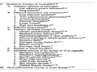
Neutrophil disorders can be grouped into deficiencies, or neutropenia, and excesses, or neutrophilia. The former can have the severe consequence of predisposing to infection, whereas the latter is usually a manifestation of an underlying inflammatory or neoplastic disease: the neutrophilia, per se, having no specific consequences. Neutropenia may reflect an inherited disease that is usually evident in childhood (such as congenital neutropenia), but it is more often acquired. The most common cause for neutropenia is the adverse effect of the use of a drug. Some cases of neutropenia have no evident cause. The health consequence of neutropenia is a function of the severity of the decrease in the blood neutrophil count and the abruptness and duration of the decrease. Qualitative disorders of neutrophils may lead to infection as a result of defective chemotaxis to an inflammatory site or defective microbial killing. Table 70-1 provides a comprehensive categorization of neutrophil disorders.


TABLE 70-1 CLASSIFICATION OF NEUTROPHIL DISORDERS


CLASSIFICATION
Table 70-1 lists disorders that result from a primary deficiency in neutrophil numbers or function. Neutropenia or neutrophilia may also occur as part of a disorder that affects multiple blood cell lineages, such as occurs in infiltrative diseases of the marrow or after cytotoxic drug therapy, but these are not included in this classification and are discussed in other parts of this text. In this classification, and in this section of the text, we consider diseases resulting from neutrophil deficiencies in which the neutrophil is either the only cell type affected or is the dominant cell type affected.
A pathophysiologic classification of neutrophil disorders has proved elusive. Techniques to measure mechanisms of impaired production or accelerated destruction of neutrophils are more difficult and complex than those used for red cells or platelets. The low concentration of blood neutrophils, accentuated in neutropenic states, makes radioactive labeling techniques to study the kinetics of autologous cells in neutropenic subjects technically difficult or not possible. The two compartments of neutrophils in the blood, the random disappearance of neutrophils from the circulation, the extremely short circulation time of neutrophils, the absence of techniques to measure the size of the tissue neutrophil compartment, and the disappearance of neutrophils by death or excretion from the tissue compartment also make multicompartment kinetic analysis exceedingly difficult. Also, neutropenic disorders are uncommon, and few laboratories are able, or prepared, to undertake the studies necessary to define the mechanisms of their development in sporadic cases. Therefore, efforts to understand the pathophysiology of neutropenia have had limited success. Hence, the classification of neutrophil disorders is partly pathophysiologic and partly descriptive (see Table 70-1). Although imperfect, classification does provide a language for communication and a basis for rectification as knowledge of the cause and mechanism of disease advances.
The classification is self-explanatory except in two areas. First, certain childhood syndromes have been listed under decreased neutrophilic granulopoiesis. They could have been listed under chronic hypoplastic or chronic idiopathic neutropenia; however, they seem to hold a special interest, and their pathogenesis is still disputed. Three childhood syndromes, although associated with neutropenia, are omitted because the neutropenia is part of a more global suppression of hemopoiesis: Pearson syndrome,1,2 Fanconi syndrome,3,4 and dyskeratosis congenita.5,6
A second area requiring explanation is the chronic idiopathic neutropenias. This group includes: (1) cases with normocellular marrows but an inadequate compensatory increase in granulopoiesis for the degree of neutropenia and (2) cases with hyperplastic granulopoiesis that is apparently ineffective. Unlike hypoplastic neutropenias in which the granulocyte precursors are markedly reduced or absent, precursors are present in the marrow in the idiopathic neutropenias, but the extent of effective granulopoiesis is probably low.
Qualitative disorders of neutrophils affect their ability to enter inflammatory exudates, to ingest microorganisms, or to kill ingested microorganisms (see Chap. 72).
CLINICAL MANIFESTATIONS
The clinical manifestations of decreased concentrations or abnormal function of neutrophils are principally the result of infection.
The combined deficit of neutrophils and monocytes characteristic of aplastic anemia, hairy-cell leukemia, and cytotoxic therapy leads to susceptibility to a broader spectrum of infectious agents. Increased concentrations of normal neutrophils per se have not been associated with clinical manifestations, although increased concentrations of leukemic neutrophil precursors can produce clinical manifestations of microcirculatory leukostasis (see Chap. 91).
NEUTROPENIA
The lower limit of the normal neutrophil count is about 1800/µl (1.8 × 10⁹/liter) in subjects of European descent and 1400/µl (1.4 × 10⁹/liter) in subjects of African descent.148,149,150,151,152,153 and 154 This finding is especially striking in Yemenite Jews, another ethnic group with very low “normal” neutrophil counts.155 A decrement in neutrophil concentration to 1000/µl (1.0 × 10⁹/liter) usually poses little threat in the otherwise healthy individual. If the neutrophil count drops further, the risk of infection increases, and subjects chronically neutropenic as a result of a production abnormality with counts less than 500 neutrophils/µl (0.5 × 10⁹/liter) are at risk of developing recurrent infections.156
The relationship of frequency or type of infection to neutrophil concentration is an imperfect one. The cause of the neutropenia, the coincidence of monocytopenia or lymphopenia, concurrent use of alcohol or glucocorticoids, and other factors can influence the likelihood of infection.
Infections in neutropenic subjects, not otherwise compromised, are most likely to result from gram-positive cocci and usually are superficial, involving skin, oropharynx, bronchi, anal canal, or vagina. However, any site may become infected, and gram-negative organisms, viruses, or opportunistic organisms may be involved.
A decrease in neutrophil count can occur abruptly or gradually (see Chap. 71). One type of drug-induced neutropenia is distinguished by the rapidity of onset. This abrupt-onset neutropenia is more likely to be severe and lead to symptoms. If the neutrophil count approaches zero (agranulocytosis), high fever; chills; necrotizing, painful oral ulcers (agranulocytic angina); and prostration may occur, presumably as a result of sepsis.157,158 and 159 As the disease progresses, headache, stupor, and rash may develop. In the preantibiotic era, persistent agranulocytosis had a fatality rate approaching 100 percent. Even with bactericidal, broad-spectrum antibiotics, severe, sustained neutropenia or agranulocytosis is a serious illness with a high fatality rate.
There is a decrease in the formation of pus in patients with severe neutropenia.160,161 This failure to suppurate can mislead the clinician and delay identification of the site of infection because minimal physical or radiographic findings develop. For example, lack of pneumonic consolidation is characteristic of pneumonia in granulocytopenic subjects. Exudate, swelling, heat, and regional adenopathy are much less prevalent in granulocytopenic patients. Fever is common, and local pain, tenderness, and erythema are nearly always present despite a marked reduction in neutrophils.162,163 and 164
The mechanism of neutropenia, as well as the severity of the deficiency of cells, plays a role in clinical manifestations. Chronic idiopathic (benign) neutropenia is associated with normal granulopoiesis in the marrow and is asymptomatic even when present for prolonged periods, sometimes in the face of neutrophil counts approaching zero.49 Presumably the delivery of neutrophils from marrow to tissues is sufficient to prevent infection despite the low blood pool size.50,51 Monocyte counts are normal, and this may also aid in host defenses, since these cells are effective phagocytes.
Chronic idiopathic (symptomatic) neutropenia is often associated with pyoderma and otitis media in children.55 The former is usually caused by Staphylococcus aureus, Escherichia coli, and Pseudomonas spp., and the latter is usually the result of infection by pneumococci or Pseudomonas aeruginosa. Unexplained chronic gingivitis also may be a manifestation of chronic neutropenia.165 Pneumonia, lung abscesses, stomatitis, hepatic abscesses, or infections in other sites may occur.56
Chronic cyclic neutropenia is characterized by periodic oscillations in the number of neutrophils, with the nadir occurring at about 3-week intervals.36,166 During neutropenia, patients develop malaise, fever, and buccal, labial, or lingual ulcers, and cervical adenopathy. Furuncles, carbuncles, cellulitis, infected cuts with lymphangitis, chronic gingivitis, and abscesses of the axilla or groin also may occur. Although severe infections may lead to fatality, life-threatening complications are uncommon (see Chap. 71).
Some individuals may have neutropenia because a larger proportion of their blood neutrophils is in the marginal rather than in the circulating pool. The total blood neutrophil pool is normal, and infections do not result from this atypical distribution of neutrophils.167 This type of alteration has been called pseudoneutropenia.
QUALITATIVE NEUTROPHIL ABNORMALITIES
Neutrophil function depends on the ability of neutrophils to adhere to endothelium, move, respond to chemotactic gradients, ingest microorganisms, and kill ingested pathogens. Loss of any of these functions can predispose to infection (see Chap. 72). Defects in each step of the neutrophil's participation in the inflammatory response have been identified.168,169 Defects in cytoplasmic contractile proteins, granule synthesis or contents, or intracellular enzymes may underlie a movement, ingestion, or killing defect. These defects may be congenital or acquired. Chronic granulomatous disease102,103 and Chédiak-Higashi disease121 are two examples of the former. Among the acquired disorders are those extrinsic to the cell, such as in the movement, chemotactic, or phagocytic defects of diabetes mellitus,168,169,170 and 171 alcohol abuse,172,173 or glucocorticoid excess.174 Acquired intrinsic disorders are usually manifestations of stem cell disorders like preleukemia175 (see Chap. 91).
Severe defects in bacterial killing, such as occur in chronic granulomatous disease, result in S. aureus, Klebsiella-Aerobacter, E. coli, and other catalase-positive bacterial infections. Suppurative lymphadenitis, pneumonia, dermatitis, hepatic abscesses, osteomyelitis, and stomatitis occur, and chronic granulomatous reactions in these sites give the disease its name. Fatality rates have been high. Functional disorders may be severe, as in chronic granulomatous disease. Mild functional disorders predispose to infections that are relatively infrequent and that respond readily to antibiotics. Severe functional disorders result in suppurative lesions because neutrophil influx into inflammatory foci is not impaired, whereas agranulocytosis is associated with nonsuppurative lesions.
NEUTROPHILIA
An overabundance of neutrophils has not been shown to result in specific clinical manifestations. Neutrophils can transiently occlude capillaries, as determined by supravital microscopy, and such occlusions may reduce local blood flow transiently and contribute to the development of ischemia.124 Impairment of reperfusion of the coronary microcirculation has been thought to be dependent, in part, on neutrophil plugging of myocardial capillaries.123
NEUTROPHIL-INDUCED VASCULAR OR TISSUE DAMAGE
Neutrophil products may contribute to the pathogenesis of inflammatory skin, bowel, synovial, glomerular, and bronchial and interstitial pulmonary diseases.124,125,126,127,128,129,130,131,132,133,134,135,136,137 and 138 In addition, these products may act as mediators of tissue injury in myocardial infarction.139,140,141 and 142 Also, highly reactive oxygen products of neutrophils may be mutagens that increase the risk of neoplasia.144,145 This action may explain, for example, the development of carcinoma of the bowel in patients with chronic ulcerative colitis and the relationship between elevated leukocyte count and the occurrence of lung cancer, independent of the effect of cigarette usage.146 The oxidants, especially hypochlorous acid and chloramines, released by the neutrophil are extremely short lived and may play a role in tissue injury by inactivating several protease inhibitors in tissue fluids, permitting proteases, especially elastase, collagenase, and gelatinase, to cause tissue injury.129 Thrombogenesis has also been ascribed to leukocyte products.143
CHAPTER REFERENCES

1.
Pearson HA, Lobel JS, Kocoshis SA, et al: A new syndrome of refractory sideroblastic anemia with vacuolization of marrow precursors and exocrine pancreatic dysfunction. J Pediatr 95:976, 1979.

2.
van de Corput MP, van den Ouweland JM, Dirks RW, et al: Detection of mitochondrial DNA deletions in human skin fibroblasts of patients with Pearson's syndrome by two-color fluorescence in situ hybridization. J Histochem Cytochem 45:55, 1997.

3.
Gordon-Smith EC, Rutherford TR: Fanconi anemia. Ballières Clin Haematol 2:139, 1989.

4.
d'Appolito M, Zelante L, Savoia A: Molecular basis of Fanconi anemia. Haematologica 83:533, 1998.

5.
Srinavin C, Trowbridge A: Dyskeratosis congenita: Clinical features and genetic aspects. J Med Genet 12:339, 1975.

6.
Dokal I: Severe aplastic anemia including Fanconi's anemia and dyskeratosis congenita. Curr Opin Hematol 3:453, 1996.

7.
Kostmann R: Infantile genetic agranulocytosis. Acta Pediatr Scand 64:362, 1975.

8.
Tidow N, Pilz C, Teichmann B, et al: Clinical relevance of point mutations in the cytoplasmic domain of granulocyte colony-stimulating factor receptor gene in patients with severe congenital neutropenia. Blood 89:2369, 1997.

9.
Rappeport JM, Parkman R, Newburger P, et al: Correction of infantile agranulocytosis (Kostmann's syndrome) by allogeneic transplantation. Am J Med 68:605, 1980.

10.
Kyds U, Pietsch T, Welte K: Expression of receptors for granulocyte colony-stimulating factor on neutrophils from patients with severe congenital neutropenia and cyclic neutropenia. Blood 79:1144, 1992.

11.
Bonilla MA, Gillio AP, Ruggeiro M, et al: Effects of recombinant human granulocyte colony-stimulating factor on neutropenia in patients with congenital agranulocytosis. N Engl J Med 320:1574, 1989.

12.
Weston B, Todd RF III, Axtell R, et al: Severe congenital neutropenia: Clinical effects and neutrophil function during treatment with granulocyte colony-stimulating factor. J Lab Clin Med 117:282, 1991.

13.
Haas RJ, Niethammer D, Goldmann SF, et al: Congenital immunodeficiency and agranulocytosis (reticular dysgenesis) Acta Paediatr Scand 66:279, 1977.

14.
Levinsky RJ, Tiedman K: Successful bone-marrow transplantation for reticular dysgenesis. Lancet 1:671, 1983.

15.
Saunders EF, Gall G, Freedman MH: Granulopoiesis in Schwachman's syndrome (pancreatic insufficiency and bone marrow dysfunction). Pediatrics 64:515, 1979.

16.
Woods WG, Roloff JS, Lukens JN: The occurrence of leukemia in patients with Schwachman syndrome. J Pediatr 99:425, 1981.

17.
Azzarà A, Carulli G, Ceccarelli M, et al: In vivo effectiveness of lithium on impaired neutrophil chemotaxis in Schwachman-Diamond syndrome. Acta Haematol 85:100, 1991.

18.
Lonsdale D, Doedhar SD, Mercer RD: Familial granulocytopenia associated with immunoglobulin abnormality. J Pediatr 71:760, 1967.

19.
Wetzler M, Talpaz M, Kleinerman ES, et al: A new familial immunodeficiency disorder characterized by severe neutropenia, a defective marrow release mechanism, and hypogammaglobulinemia. Am J Med 89:663, 1990.

20.
Kozlowski C, Evans DIK: Neutropenia associated with X-linked agammaglobulinemia. J Clin Pathol 44:388, 1991.

21.
Lux SE, Johnston RB Jr, August CS, et al: Chronic neutropenia and abnormal cellular immunity in cartilage-hair hypoplasia. N Engl J Med 282:231, 1970.

22.
Trojak JE, Polmar SH, Winkelstein JA: Immunologic studies of cartilage-hair hypoplasia in the Amish. Johns Hopkins Med J 148:157, 1981.

23.
Norio R, Raitta C, Lindahl E: Further delineation of the Cohen syndrome. Clin Genet 25:1, 1984.

24.
Warburg M, Pedersen SA, Hønlyk H: The Cohen syndrome. Ophthalmic Pediatr Genet II:7, 1990.

25.
Barth PG, Scholte HR, Berden JA, et al: An X-linked mitochondrial disease affecting cardiac muscle, skeletal muscle and neutrophil leukocytes. J Neurol Sci 62:327, 1983.

26.
Bohurs PA, Hensels GW, Hulsebos TJM, et al: Mapping of the locus for the X-linked cardioskeletal myopathy with neutropenia and abnormal mitochondria (Barth syndrome) to Xq28. Am J Hum Genet 48:481, 1991.

27.
Bassan R, Viero P, Minetti B, et al: Myelokathexis: A rare form of chronic benign neutropenia. Br J Haematol 58:115, 1984.

28.
Wetzler M, Talpaz M, Kellagher MJ, et al: Myelokathexis. JAMA 267:2179, 1992.

29.
Lightsey AL, Parmley RT, Marsh WL, et al: Severe congenital neutropenia with unique features of dysgranulopoiesis. Am J Hematol 18:59, 1985.

30.
Koenig JM, Christensen RD: Incidence, neutrophil kinetics and natural history of neonatal neutropenia associated with maternal hypertension. N Engl J Med 321:557, 1989.

31.
Koenig JM, Christensen RD: The mechanism responsible for diminished neutrophil production in neonates delivered of women with pregnancy-induced hypertension. Am J Obstet Gynecol 165:467, 1991.

32.
Hartl PW: Drug-induced agranulocytosis, in Blood Disorders Due to Drugs and Other Agents, edited by RH Girdwood, pp 147–186. Excerpta Medica, Amsterdam, 1974.

33.
Hine LK, Gerstman BB, Wise RP, Tsang Y: Mortality resulting from blood dyscrasias in the United States, 1984. Am J Med 88:151, 1990.

34.
Pisciotta AV: Drug-induced agranulocytosis peripheral destruction of polymorphonuclear leukocytes and their marrow precursors. Blood Rev 4:226, 1990.

35.
Julia A, Olona M, Bueno J, et al: Drug-induced agranulocytosis. Br J Haematol 79:366, 1991.

36.
Wright DG, Dale DC, Fauci AS, Wolff SM: Human cyclic neutropenia. Medicine 60:13, 1981.

37.
Tefferi A, Solberg LA, Petett RM, Willis LG: Adult-onset cyclic bicytopenia. Am J Hematol 30:181, 1989.

38.
Loughran TP Jr, Clark EA, Hammond WP: Adult-onset cyclic neutropenia is associated with increased large granular lymphocytes. Blood 68:1082, 1986.

39.
Hammond WP, Price TH, Souza LM, Dale DC: Treatment of cyclic neutropenia with granulocyte colony-stimulating factor. N Engl J Med 320:1306, 1989.

40.
Marinone G, Roncoli B, Marinone MG: Pure white cell aplasia. Semin Hematol 28:298, 1991.

41.
Morley AA, Carew JP, Baikie AG: Familial cyclic neutropenia. Br J Haematol 13:719, 1967.

42.
Hammond WP, Chatta GS, Andrews RG, Dale DC: Abnormal responsiveness of granulocyte-committed progenitor cells in cyclic neutropenia. Blood 79:2536, 1992.

43.
Dale DC, Hammond WP: Cyclic neutropenia: A clinical review. Blood Rev 2:178, 1988.

44.
Spaet TH, Dameshek W: Chronic hypoplastic neutropenia. Am J Med 13:35, 1952.

45.
Hutchinson R, Bunnell K, Thorne J: Suppression of granulopoietic progenitor cell proliferation by metabolites of the branched-chain amino acids. J Pediatr 106:62, 1985.

46.
Murdock JMC, Smith CC: Haematologic aspects of systemic disease—infection. Clin Haematol 1:619, 1972.

47.
Olson JP, Lichtman MA: Neutropenia, in Hematology for Practitioners, edited by MA Lichtman, pp 105–120. Little, Brown, Boston, 1978.

48.
Cutting HO, Lange JE: Familial-benign chronic neutropenia. Ann Intern Med 61:876, 1964.

49.
Kyle RA: Natural history of chronic idiopathic neutropenia. N Engl J Med 302:908, 1970.

50.
Wright DG, Meierovics AI, Foxley JM: Assessing the delivery of neutrophils to tissues in neutropenia. Blood 67:1023, 1986.

51.
Mant MJ, Gordon PA, Akabotu JJ: Bone marrow granulocyte reserve in chronic benign idiopathic neutropenia. Clin Lab Haematol 9:281, 1987.

52.
Logue GL, Shastri KA, Laughlin M, et al: Idiopathic neutropenia: Antineutrophil antibodies and clinical correlations. Am J Med 90:211, 1991.

53.
Jonsson OG, Buchanan GR: Chronic neutropenia during childhood. Am J Dis Child 145:232, 1991.

54.
Jakubowski AA, Souza L, Kelly F, et al: Effects of human granulocyte colony-stimulating factor in a patient with idiopathic neutropenia. N Engl J Med 320:38, 1989.

55.
Pincus SH, Boxer LA, Stossel TP: Chronic neutropenia in childhood. Am J Med 61:849, 1976.

56.
Dale DC, Guerry D IV, Wewerka JR, et al: Chronic neutropenia. Medicine 58:128, 1979.